 |
Clinical Image
An epidermoid cyst of the submandibular space
1 Senior Registrar Otolaryngology, Department of Clinical Surgical Sciences, University of West Indies, Eric Williams Medical Sciences Complex, Champs Fleur, Trinidad and Tobago
2 Consultant Otolaryngologist, Department of Clinical Surgical Sciences, University of West Indies, Eric Williams Medical Sciences Complex, Champs Fleur, Trinidad and Tobago
Address correspondence to:
Nicholas Figaro
Department of Surgery, Eric Williams Medical Sciences Complex, Uriah Butler Highway, Champ Fleurs,
Trinidad and Tobago
Message to Corresponding Author
Article ID: 100005Z18NF2022
Access full text article on other devices

Access PDF of article on other devices


How to cite this article
Figaro N, Ramoutar R, Arozarena R, Juman S. An epidermoid cyst of the submandibular space. J Case Rep Images Otolaryngol 2022;3(2):1–3.ABSTRACT
No Abstract
Keywords: Cervical cyst, Epidermoid cyst, Submandibular space
Case Report
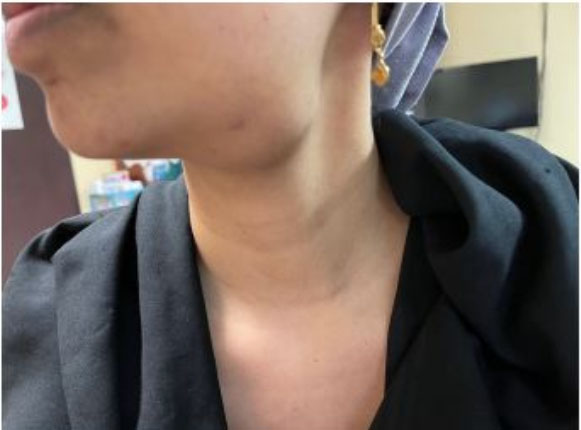
A 35-year-old female presented with a 6-month history of a mass in the left submandibular region. Despite being asymptomatic, the patient became concerned with the cosmesis of the progressively enlarging lesion. She did not indicate any antecedent history of head and neck infection, trauma, or surgery. Physical examination revealed a 3.0 × 4.0 cm soft, non-tender, mobile mass in the left submandibular region (Figure 1). The mass was not attached to the overlying skin and the patient’s oral cavity examination was normal.
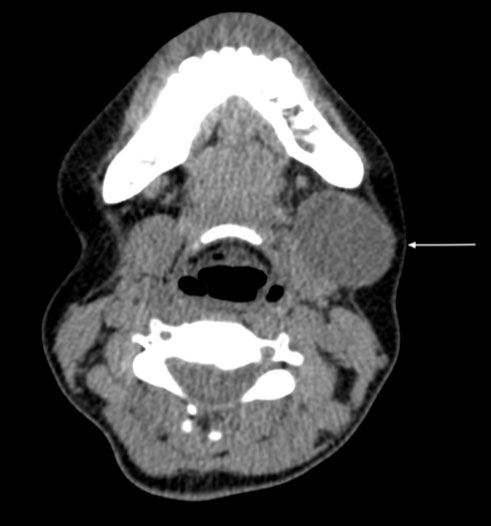
Contrast enhanced computed tomography imaging demonstrated a well-defined cystic lesion measuring 3.5 × 2.8 cm, positioned under the platysma and arising from the antero-lateral surface of the left submandibular gland, displacing the submandibular gland posteriorly (Figure 2). These findings led to a radiological diagnosis of a dermoid cyst of the submandibular gland. Fine needle aspiration cytology revealed an admixture of degenerate squamous cell and keratinous debris suggestive of an epidermoid cyst.
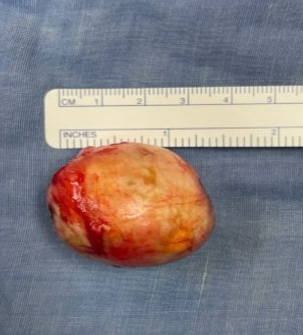
Surgical excision was performed under general anesthesia. A transverse skin incision was made parallel to the lower border of the mandible below the lesion. Careful dissection facilitated removal of the lesion in toto, with no disruption of the submandibular gland (Figure 3). The patient had an uneventful post-operative period with no recurrence after 1 year.
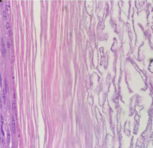
Gross pathological assessment revealed a unilocular cyst containing yellow creamy material, with no skin adnexal elements. Histopathological analysis revealed a thin-walled cyst lined by stratified squamous epithelium with an attenuated granular layer, lamellated keratin, and keratinaceous debris. These features are consistent with the diagnosis of an epidermoid cyst (Figure 4).
Discussion
Epidermoid and dermoid cysts are uncommonly found in the head and neck region. They account for approximately 7% of all benign swellings of the head and neck with less than 2% occurring in the oral cavity [1]. An epidermoid cyst is defined as a simple cystic lesion composed of keratinized squamous epithelium containing semi-solid epithelial debris without any skin appendages [2]. Whereas a dermoid cyst has skin adnexa like hair, hair follicles, sebaceous, and sweat glands that are enclosed within its epithelial lining [3].
Epidermoid cysts are a result of epithelial tissue aberration in the dermis secondary to traumatic implantation or congenitally derived from entrapped residual ectodermal tissue during embryogenesis [4]. Dermoid cysts, although they are formed similarly by entrapped embryonic ectodermal remnants, they are exclusively found in areas of fusion, as such, they are typically located in the midline or sublingual region of the oral cavity [4].
Several cases of epidermoid cysts located in the submandibular region were previously described; however, they were generally found within the mylohyoid and hyoglossus muscles, medial to the submandibular gland [3],[5],[6]. This suggests progressive lateral migration from the initial midline region of the floor of the mouth where the cysts developed. Pericystic adhesions and cord-like constructs are also suggestive of cystic migration [3].
This report describes a unique case of an epidermoid cyst found on the lateral surface of the submandibular gland. We believe that this epidermoid cyst may have been congenital in origin, based on the patient’s history of no trauma, inflammation, or surgery in the submandibular region and no intraoperative findings of adhesions or any cord-like structural attachments.
The differential diagnoses for submandibular swellings are extensive, ranging from benign pathologies like sialadenitis, sialolithiasis, branchial cysts, and lipomas to malignant lesions like primary submandibular gland tumors, metastatic cervical lymph nodes, and lymphomas [7]. The plethora of submandibular pathologies makes preoperative diagnosis essential for definitive management of the patient [2],[4]. In our case, radiological imaging and fine needle aspiration cytology were highly suggestive of an epidermoid cyst for which complete surgical excision is required for both a definitive diagnosis and a cure. Meticulous dissection is required to prevent inadvertent injury to the mandibular branch of the facial nerve, lingual nerve, and hypoglossal nerve, as well as spillage of cystic contents into the wound causing postoperative inflammation [2],[5]. The frequency of recurrence and malignant transformation of an epidermoid cyst is nominal [1],[4],[7].
Conclusion
An epidermoid cyst of the submandibular space is an unusual entity and can pose a diagnostic dilemma due to the pathologic diversity that can occur at this anatomical subsite. Definitive diagnosis and treatment is achieved with complete surgical excision of the lesion.
REFERENCES
1.
Janarthanam J, Mahadevan S. Epidermoid cyst of submandibular region. J Oral Maxillofac Pathol 2012;16(3):435–7. [CrossRef]
[Pubmed]

2.
Patil N, Shukla D, Sonawane R. A rare case of epidermal cyst of the submandibular gland. International Journal of Health Sciences and Research 2016;6(6):434–6.
3.
Rosen D, Wirtschafter A, Rao VM, Wilcox TO Jr. Dermoid cyst of the lateral neck: A case report and literature review. Ear Nose Throat J 1998;77(2):125– 32.
[Pubmed]
4.
Kudoh M, Harada H, Omura K, Ishii Y. Epidermoid cyst arising in the submandibular region. Case Rep Med 2013;2013:419289. [CrossRef]
[Pubmed]
5.
Prepageran N, Rahmat O, Kuljit S. Epidermal cyst of submandibular gland. Med J Malaysia 2005;60(4):483–4.
[Pubmed]
6.
Dutt SN, Hock YL, Saleem Y, Bhat N, East DM. Epidermoid cyst of the submandibular gland. Indian J Otolaryngol Head Neck Surg 2000;52(4);378–9. [CrossRef]
[Pubmed]
7.
Kar R, Thorawade V, Jagade M, et al. An unusal case of epidermal cyst in submandibular space. International Journal of Otolaryngology and Head & Neck Surgery 2014;3(5):213–5. [CrossRef]
SUPPORTING INFORMATION
Author Contributions
Nicholas Figaro - Conception of the work, Design of the work, Acquisition of data, Analysis of data, Drafting the work, Revising the work critically for important intellectual content, Final approval of the version to be published, Agree to be accountable for all aspects of the work in ensuring that questions related to the accuracy or integrity of any part of the work are appropriately investigated and resolved.
Rickhi Ramoutar - Acquisition of data, Drafting the work, Final approval of the version to be published, Agree to be accountable for all aspects of the work in ensuring that questions related to the accuracy or integrity of any part of the work are appropriately investigated and resolved.
Rodolfo Arozarena - Analysis of data, Revising the work critically for important intellectual content, Final approval of the version to be published, Agree to be accountable for all aspects of the work in ensuring that questions related to the accuracy or integrity of any part of the work are appropriately investigated and resolved.
Solaiman Juman - Analysis of data, Revising the work critically for important intellectual content, Final approval of the version to be published, Agree to be accountable for all aspects of the work in ensuring that questions related to the accuracy or integrity of any part of the work are appropriately investigated and resolved.
Guaranter of SubmissionThe corresponding author is the guarantor of submission.
Source of SupportNone
Consent StatementWritten informed consent was obtained from the patient for publication of this article.
Data AvailabilityAll relevant data are within the paper and its Supporting Information files.
Conflict of InterestAuthors declare no conflict of interest.
Copyright© 2022 Nicholas Figaro et al. This article is distributed under the terms of Creative Commons Attribution License which permits unrestricted use, distribution and reproduction in any medium provided the original author(s) and original publisher are properly credited. Please see the copyright policy on the journal website for more information.


