 |
Case Report
Dermoid cyst presenting as a submental swelling, causing diagnostic confusion
1 Department of ENT, Al Kuwait Hospital, Dubai, UAE
2 Department of Pathology, Al Qassimi Hospital, Sharjah, UAE
Address correspondence to:
Mohammed Sadath
Department of ENT, Al Kuwait Hospital, Dubai,
UAE
Message to Corresponding Author
Article ID: 100010Z18MS2023
Access full text article on other devices

Access PDF of article on other devices


How to cite this article
Sadath M, Al Amadi A, Busafared FA, Alshaiba AA, Idnani DD. Dermoid cyst presenting as a submental swelling, causing diagnostic confusion. J Case Rep Images Otolaryngol 2023;4(2):6–9.ABSTRACT
Dermoid cyst is a benign cutaneous developmental anomaly that arises from the entrapment of ectodermal elements commonly arising from midline where the embryonic structures fuse. It is lined by stratified squamous epithelium with mature skin appendages found on their wall and their lumens filled with keratin and hair. Dermoid cysts usually form in the ovary, testes, the skin of the head, neck, face, or lower back, or in the central nervous system. Rarely dermoid cyst can occur on the lateral part of the neck or floor of the mouth. Neck swellings are difficult to diagnose clinically and are close to vital structures. Radiological investigations will help in diagnosis, but it is not confirmatory. We report a case of a 9-year-old girl who attended our institution with a right-sided neck swelling that had been present for five years and had an occasional history of pain. Examination of neck showed submental swelling above the hyoid, toward the right side, 3 × 4 cm, soft cystic, nontender, moving with deglutition and protrusion of tongue. Intraorally, the swelling was not visible, but it was bimanually palpable. Treated surgically and excision biopsy reported as a dermoid cyst. In the present article we report this rare mode of presentation of a dermoid cyst with a diagnostic confusion and a surgical challenge.
Keywords: Dermoid cyst, Floor of mouth, Histopathology, Ranula, Thyroglossal cyst
Introduction
Dermoid cyst is a benign cutaneous developmental anomaly that arises from the entrapment of ectodermal elements commonly arising from midline where the embryonic structures fuse [1],[2],[3]. Dermoid cysts usually form in the ovary, testes, the skin of the head, neck, face, or lower back, or in the central nervous system. Rarely dermoid cyst can occur on the lateral part of the neck or floor of the mouth.
A 9-year-old girl attended our institution with a right-sided neck swelling that had been present for five years and had an occasional history of pain. Examination of the neck showed submental swelling above the hyoid, toward the right side. It was soft, cystic, nontender, and moving with deglutition and tongue protrusion. The possible differential diagnosis was thyroglossal duct cyst, ranula or less likely dermoid cyst [4],[5]. The patient underwent surgical excision and biopsy revealed a dermoid cyst [6].
Case Report
A 9-year-old girl attended our institution with a right-sided neck swelling that had been present for five years and has an occasional history of pain. Examination of neck showed submental swelling above the hyoid, toward right side, 3 × 4 cm, soft cystic, nontender, moving with deglutition and protrusion of tongue. Intraorally, the swelling was not visualized, but it was bimanually palpable. Ear, nose, and throat examination was normal.
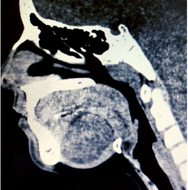
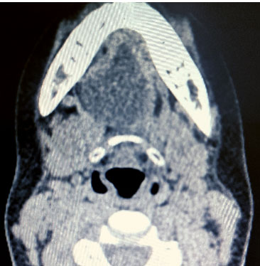
Ultrasound scan of the neck revealed a well-demarcated cyst, 35 mm × 24 mm in size, with a normal smooth avascular thin wall, reported as a thyroglossal cyst. Computed tomography (CT) of soft tissue neck showed a hypodense, cystic lesion in submandibular location slightly to the right, 36 mm × 27 mm × 39 mm (Figure 1 and Figure 2). The inferior extent of the lesion was at the level of the hyoid bone, and it extended up to the floor of the mouth. Anteriorly it was reaching up to the base of the mandible and posteriorly up to the hyoid bone. Reported probable differential diagnosis were, thyroglossal duct cyst, ranula or less likely dermoid cyst.
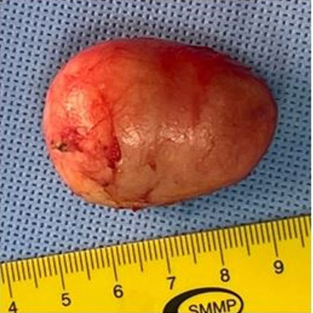
After detailed clinical and radiological workup, surgical exploration and excision of cyst was done under General Anesthesia. During the surgical procedure, submental swelling, toward the right side, deep to the Mylohyoid muscle, 4.5 cm × 3 cm in size, nodular, soft, cystic, completely encapsulated mass was dissected out and sent for histopathology (Figure 3).
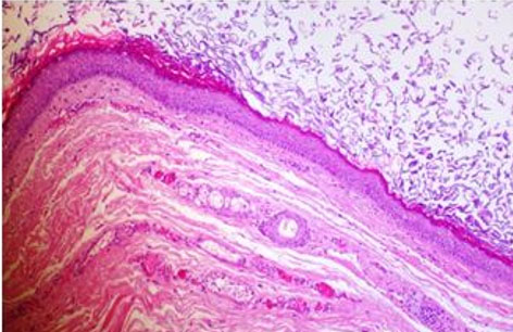
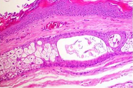
Histopathology showed cyst wall lined by stratified squamous epithelium with granular layer and mature skin appendages (hair follicles and sebaceous glands) loose keratin flakes in the lumen, reported as dermoid cyst (Figure 4 and Figure 5).
The patient reviewed in outpatient clinic after 10 days and on examination of the neck, the incision site was fully healed.
Discussion
Dermoid cysts are developmental lesions found inside normal organs as a result of the inclusion of tissue from diverse sources, such as ectoblastic, mesoblastic, or endoblastic origin [1],[2],[3]. Dermoid cyst in neck is rare and that occurs submentally in floor of mouth, not confined to midline is extremely rare. It is caused by a defect in the fusion of the embryonic lateral mesenchymatic mass, mainly the first and second pharyngeal arches during the 5th week of embryonic development [1].
There are three histological variants, the true dermoid cyst, the epidermoid cyst, and the teratoid cyst [1]. True dermoid cysts lined with epithelium with keratinization and identifiable skin on the cyst wall. Epidermoid cysts are lined with simple squamous epithelium with a fibrous wall. Teratoid cyst is lined with simple squamous and ciliated respiratory epithelium that contains derivatives of ectoderm, mesoderm, and endoderm. In all three histological variants, cyst usually filled with thick, greasy material.
Dermoid cysts of neck commonly appear as a painless, slow-growing mass in anterior neck towards the midline [1],[3],[7]. Lateral presentation is rare. Clinical examination showing a soft or firm, nontender swelling. While clinically evaluating present case, it was moving with deglutition and tongue protrusion, so possibility of thyroglossal cyst could not rule out, and ultrasound report was also supported thyroglossal cyst. It was bimanually palpable soft cystic mass; hence, Ranula was another differential diagnosis. Although radiological investigations were not diagnostic, they provided a clue to the diagnosis and guided us during surgery regarding the extent of the lesion [4],[5]. Surgical excision and biopsy is diagnostic and curative.
Surgical approach depends on size of the cyst and its relation to muscles of the neck and oral cavity. Options are intraoral and extraoral. In intraoral approach the exposure is limited and there is risk of injury to lingual and hypoglossal nerves [2]. Extraoral approach is the most suitable option for large-sized cysts that cross the midline and provide good exposure [6].
Conclusion
Submental dermoid cysts, not confined to the midline, are very rare. However, we should consider them as a differential diagnosis. Although radiological investigations like ultrasound scan and CT scan were not diagnostic, but provided a clue to the diagnosis, and helped us to assess the extent of mass and to plan the surgical approach. The final diagnosis is achieved by surgical exploration and excision biopsy.
REFERENCES
1.
Makos C, Noussios G, Peios M, Gougousis S, Chouridis P. Dermoid cysts of the floor of the mouth: Two case reports. Case Rep Med 2011;2011:362170. [CrossRef]
[Pubmed]

2.
Somashekhar SN, Kulkarni A. Dermoid cyst of neck: A diagnostic dilemma and a therapeutic challenge. Int J Head Neck Surg 2017;8(3):125–7. [CrossRef]
3.
Saheeb BDO, Osaguona A. Submental dermoid cyst: A case report. OJM 2005;17(1&2):24–7. [CrossRef]
4.
Celenk P, Celenk C, Kocasarac HD. Imaging features of sublingual dermoid cysts: A report of four cases. Radiol Case Rep 2022;17(8):2888–93. [CrossRef]
[Pubmed]
5.
Sanal HT. Ultrasound finding of dermoid cysts located in neck region: Sack-of-Marble sign. KBB - Forum Electronic Journal of Otolaryngology - Head and Neck Surgery 2008;7(1).
6.
Regis DM, Cunha JLS, Sánchez-Romero C, da Cruz Ramos MAC, de Albuquerque RLC, Bezerra BT. Diagnosis, management, and follow-up of extensive dermoid cyst of the submental region. Autops Case Rep 2019;9(3):e2019095. [CrossRef]
[Pubmed]
7.
Das A, Trupthi MC, War SS, Varghese AM. Paediatric submental epidermoid cyst. BMJ Case Rep 2022;15(7):e250722. [CrossRef]
[Pubmed]
SUPPORTING INFORMATION
Author Contributions
Mohammed Sadath - Conception of the work, Design of the work, Acquisition of data, Analysis of data, Drafting the work, Revising the work critically for important intellectual content, Final approval of the version to be published, Agree to be accountable for all aspects of the work in ensuring that questions related to the accuracy or integrity of any part of the work are appropriately investigated and resolved.
Ahmad Al Amadi - Analysis of data, Revising the work critically for important intellectual content, Final approval of the version to be published, Agree to be accountable for all aspects of the work in ensuring that questions related to the accuracy or integrity of any part of the work are appropriately investigated and resolved.
Faiza Ahmed Busafared - Analysis of data, Drafting the work, Final approval of the version to be published, Agree to be accountable for all aspects of the work in ensuring that questions related to the accuracy or integrity of any part of the work are appropriately investigated and resolved.
Aisha Abdulatif Alshaiba - Analysis of data, Drafting the work, Final approval of the version to be published, Agree to be accountable for all aspects of the work in ensuring that questions related to the accuracy or integrity of any part of the work are appropriately investigated and resolved.
Desh Deepak Idnani - Acquisition of data, Drafting the work, Final approval of the version to be published, Agree to be accountable for all aspects of the work in ensuring that questions related to the accuracy or integrity of any part of the work are appropriately investigated and resolved.
Guaranter of SubmissionThe corresponding author is the guarantor of submission.
Source of SupportNone
Consent StatementWritten informed consent was obtained from the patient for publication of this article.
Data AvailabilityAll relevant data are within the paper and its Supporting Information files.
Conflict of InterestAuthors declare no conflict of interest.
Copyright© 2023 Mohammed Sadath et al. This article is distributed under the terms of Creative Commons Attribution License which permits unrestricted use, distribution and reproduction in any medium provided the original author(s) and original publisher are properly credited. Please see the copyright policy on the journal website for more information.


