 |
Case Report
A case of an oncocystic laryngeal cyst: A rare pathology
1 Wagga Wagga Base Hospital, Wagga Wagga, NSW, Australia
2 UNSW Sydney, Sydney, Australia
Address correspondence to:
Madison Boot
27/3 King Street, Newcastle, NSW 2300,
Australia
Message to Corresponding Author
Article ID: 100012Z18MB2024
Access full text article on other devices

Access PDF of article on other devices


How to cite this article
Boot M, Lathif A. A case of an oncocystic laryngeal cyst: A rare pathology. J Case Rep Images Otolaryngol 2024;5(2):1–3.ABSTRACT
Introduction: Oncocytic cysts of the larynx are rare benign lesions characterized by slow growth and large polygonal oncocytic cells. They are primarily found in the supraglottic area and ventrals, with a prevalence of 0.5–1% in laryngeal biopsies.
Case Report: A 75-year-old female presented with progressive dysphonia for eight months, with no other significant symptoms. Examination revealed a pedunculated polypoidal mass on the right false vocal cord. Imaging confirmed a well-defined lesion. Microlaryngoscopy and excision biopsy revealed an oncocytic cyst, leading to symptom improvement post-surgery.
Conclusion: Oncocytic cysts are often mistaken for Warthin tumors histologically but lack bilayered oncocytes and lymphoid stroma. They are associated with aging, chronic inflammation, and possibly smoking. Clinical presentation typically includes chronic hoarseness, and management involves local excision and follow-up due to potential recurrence. This case highlights the rare occurrence of oncocytic cysts in the larynx, emphasizing the importance of considering this diagnosis in patients presenting with hoarseness. Proper histological evaluation and management can lead to symptom resolution and favorable outcomes.
Keywords: Dysphonia, Laryngeal neoplasm, Oncocytic cyst, Oncocytes
Introduction
Oncocytic cysts of the larynx are benign, slow-growing large polygonal oncocytic cells often involving the supraglottic area of the false vocal cords and the ventrals [1]. They were first described in 1946 by pathologist Dr. H. Nohteri [2]. They are identified by the large numbers of hypertrophied mitochondria producing eosinophilia and a swollen appearance [2]. Oncocytic cysts of the larynx are rare, representing 0.5–1% of all laryngeal mass biopsies, and to date, approximately 155 cases have been reported [3]. Herein, we discuss a rare case of an oncocytic cyst diagnosed in a 75-year-old female.
Case Report
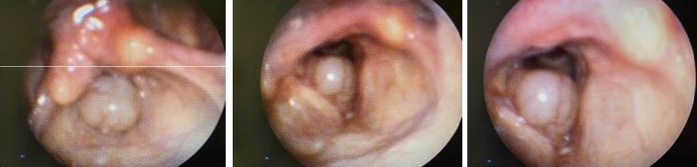
A 75-year-old female presented to an otolaryngology clinic with an eight-month history of worsening hoarseness of voice. The hoarseness of voice was constant and associated with occasional postnasal drip. There was no history of difficulty breathing, dyspnea, stridor, weight loss, night sweats, or throat clearing. Her past medical history included reflux, and emphysema. There was no personal or family history of malignancy. The patient was a smoker of 60 pack-years and had minimal alcohol intake. On examination, her oropharynx, oral, and neck examination was unremarkable. Flexible laryngoscopy showed a large pedunculated polypoidal mass from the right false vocal cord, which flipped into the rima glottidis during phonation, which can be seen in Figure 1. Vocal cords moved symmetrically and were otherwise of normal appearance. Computed tomography (CT) of the chest and neck showed a 10 × 11 × 11 mm low attenuation, non-enhancing well-defined lesion arising from the anterior aspect of the right vocal cord. There were no enlarged cervical lymph nodes.
The decision was made to proceed with microlaryngoscopy and excision biopsy of the right vocal cord lesion. Intraoperatively there was a large cystic mass from the right ventricle with normal-appearing vocal cords. The lesion was excised with clear margins. Histopathology identified a cystic cavity containing mucoid secretions lined by the pseudostratified oncocytic columnar epithelium of varying thickness with no evidence of malignancy. These features were in keeping with an oncocytic cyst.
Postoperatively, the patient recovered well with almost an immediate improvement in hoarseness of voice. Six months postoperatively, the patient was symptom-free and flexible and showed no residual or recurrence of the growth. The patient was organized for a follow-up with an otolaryngologist in 12 months.
Discussion
Oncocytic cysts of the larynx are also known as oncocytoma, oncocytic papillary cystadenoma, papillary cystadenoma, and oxyphilic granular cells adenoma throughout literature. These lesions are slow-growing, benign lesions of that most commonly occur in the upper respiratory tract. They rarely occur in the larynx and account for 0.5–1% of laryngeal biopsies [3]. The first case of an oncocytic laryngeal cyst was reported in 1946 by pathologist Dr. H. Nohteri; since then, roughly 155 cases have been reported [2],[3].
Laryngeal oncocytic cysts are often found in the elderly and females. The exact pathophysiology is unknown; however, the metaplastic process is thought to be caused by a high level of metabolic activity, inflammation, degenerative process, and cell aging [3],[4],[5],[6]. It is often associated with increased activity of respiratory chain enzymes such as succinate dehydrogenase and cytochrome c-oxidase, suggesting tissue response to chronic irritation and perhaps associated with cigarette smoking [3],[4]. The most common site of occurrence within the larynx is the supraglottic area of the false vocal cords and the ventrals. This is thought to be due to the large amounts of glandular tissue in the lamina propria [3],[4].
Dysphonia is the most common presenting symptoms. The dysphonia can be further classified as hoarseness which is often chronic and persists for weeks to years before diagnosis [1]. Patients may also present with dyspnea, chronic cough, and globus sensation. Rarely do these cysts present with pain, stridor, or laryngeal obstruction, in which cases these are late signs of disease. There has been one reported case of oncocytic laryngeal cyst resulting in acute vocal cord paralysis and sudden respiratory death [7].
Investigations commonly used to assess and diagnose patients with oncocytic cysts include direct visualisation with flexinasoendoscopy, common forms of imaging modality such as computed tomography (CT) or magnetic resonance imaging (MRI) and tissue diagnosis. The most common appearance of an oncocytic cyst on flexinasoendoscopy is a well circumscribed round well demarcated pedunculated cystic lesion extending from the supraglottic structures. Oncocytic cysts are commonly described as a well-defined, fluid-attenuation or fluid signal intense, non-enhancing rounded lesion on CT and MRI [8].
Formal diagnosis can be made with tissue sampling and histopathological testing. Oncocytic cysts of the larynx are slow growing large polygonal oncocytic cells. Histologically they are lined predominately or exclusively by onococytes, large polygonal cells with hyperchromatic nuclei and eosinophilic granular cytoplasm [4]. Due to the microscopic similarities may be mistaken for a Warthin tumor, however, they can be distinguished by the lack of bilayer oncocytes and lymphoid stroma [5].
Recommended management of oncocytic cysts of the larynx included local excision and postoperative follow-up. Local excision can vary from complete excision through laryngofissure to endoscopic laser excision or marsupialization with carbon dioxide laser. Once diagnosed with an oncocytic cyst associated recurrence is high and therefore routine follow-up is recommenced. Oncocytic cysts are benign lesions and there are no definite reports of malignant transformation association with squamous carcinoma of the larynx has been reported [3],[4],[5].
Specifically, in the review of our case, our patient was an elderly female, consistent with the epidemiological factors often seen in these cases. Our patient also had a significant smoking history which may have been a contributing factor to this presentation. The presenting symptom was typical for this disease, and her treatment management was performed per recommended guidelines. Postoperative follow-up has shown no recurrences; however, the patient will continue to be reviewed to assess for disease recurrence.
Conclusion
In conclusion, this case underscores the importance of considering oncocytic cysts in the differential diagnosis of laryngeal lesions, particularly in elderly patients presenting with chronic dysphonia. Proper histopathological assessment is crucial for accurate diagnosis and appropriate management, which often involves surgical excision and diligent postoperative follow-up due to the potential for recurrence. Understanding the histological features, clinical presentation, and management strategies for oncocytic cysts contributes to improved patient outcomes and highlights the need for ongoing research to enhance our understanding of this rare entity.
REFERENCES
1.
Salerno G, Mignogna C, Cavaliere M, D’Angelo L, Galli V. Oncocytic cyst of the larynx: An unusual occurrence. Acta Otorhinolaryngol Ital 2007;27(4):212–5.
[Pubmed]

2.
Nohteri H. A case of laryngeal cyst composed of oncocytes and the appearance of oncocytes in the mucous membrane of the nose and the larynx. Acta Pathologica Microbiologica Scandinavica 2009;23(6):473–83.
3.
Heyes R, Tomblinson CM, Lott DG. Multiple and recurrent oncocytic cysts of the larynx. Ear Nose Throat J 2020;99(5):NP54–5. [CrossRef]
[Pubmed]
4.
Mettias B, Usanov A, Osborne J. Oncocytic cyst of the larynx: Precipitating factors and mode of treatment (case report). Egyptian Journal of Ear, Nose, Throat and Allied Sciences 2012;13(2):91–3.
5.
Ferlito A, Recher G. Oncocytic lesions of the larynx. Arch Otorhinolaryngol 1981;232(2):107–15. [CrossRef]
[Pubmed]
6.
Dhingra JK, Aqel NM, McEwen J, Bleach NR. Multiple oncocytic cysts of the larynx. J Laryngol Otol 1995;109(12):1226–8. [CrossRef]
[Pubmed]
7.
Westerberg BD, Durham JS, Berean KW. Multiple oncocytic laryngeal cysts presenting as acute airway obstruction. J Otolaryngol 1995;24(5):319–21.
[Pubmed]
8.
DeSanto LW, Devine KD, Weiland LH. Cysts of the larynx—classification. Laryngoscope 1970;80(1):145–76. [CrossRef]
[Pubmed]
SUPPORTING INFORMATION
Author Contributions
Madison Boot - Conception of the work, Design of the work, Acquisition of data, Analysis of data, Drafting the work, Revising the work critically for important intellectual content, Final approval of the version to be published, Agree to be accountable for all aspects of the work in ensuring that questions related to the accuracy or integrity of any part of the work are appropriately investigated and resolved.
Abdul Lathif - Conception of the work, Design of the work, Acquisition of data, Analysis of data, Drafting the work, Revising the work critically for important intellectual content, Final approval of the version to be published, Agree to be accountable for all aspects of the work in ensuring that questions related to the accuracy or integrity of any part of the work are appropriately investigated and resolved.
Guaranter of SubmissionThe corresponding author is the guarantor of submission.
Source of SupportNone
Consent StatementWritten informed consent was obtained from the patient for publication of this article.
Data AvailabilityAll relevant data are within the paper and its Supporting Information files.
Conflict of InterestAuthors declare no conflict of interest.
Copyright© 2024 Madison Boot et al. This article is distributed under the terms of Creative Commons Attribution License which permits unrestricted use, distribution and reproduction in any medium provided the original author(s) and original publisher are properly credited. Please see the copyright policy on the journal website for more information.


