 |
Case Report
Tonsillar lipoma: A case report from Saudi Arabia
1 Otolaryngology Registrar, Department of Otolaryngology – Head & Neck Surgery, Prince Sultan Military Medical City, Riyadh, Saudi Arabia
2 Otolaryngology consultant, Department of Otolaryngology Head & Neck Surgery, King Fahad Military Medical City, Saudi Arabia
Address correspondence to:
Lenah Khalid AlFadhil
MD, Otolaryngology Registrar, Department of Otolaryngology – Head & Neck Surgery, Prince Sultan Military Medical City, Riyadh,
Saudi Arabia
Message to Corresponding Author
Article ID: 100016Z18LF2025
Access full text article on other devices

Access PDF of article on other devices


How to cite this article
AlFadhil L, AlGhamdi AM. Tonsillar lipoma: A case report from Saudi Arabia. J Case Rep Images Otolaryngol 2025;4(2):5–8.ABSTRACT
Introduction: Lipoma is one of the rarest benign tumors affecting the tonsils, with very few cases reported in the literature. Due to its rarity, it can be easily overlooked in the differential diagnosis of tonsillar masses.
Case Report: We report a case of a 28-year-old Saudi male who presented with a foreign body sensation in the throat. Clinical examination revealed a mass arising from the upper pole of the right tonsil. The patient underwent bilateral tonsillectomy, and histopathological analysis confirmed the diagnosis of lipoma. No recurrence was observed at the three-month follow-up.
Conclusion: Tonsillar lipomas are uncommon and may present with nonspecific symptoms. This case highlights the importance of considering lipoma in the differential diagnosis of unilateral tonsillar enlargement and discusses the rationale for surgical management in such rare presentations.
Keywords: Benign tonsillar tumor, Bilateral tonsillectomy, Case report, Foreign body sensation, Tonsillar lipoma
Introduction
Lipoma is a benign neoplasm composed of abnormal adipose tissue, most commonly found in subcutaneous regions [1]. Although lipomas are the most frequent mesenchymal tumors, only approximately 15% arise in the head and neck region, affecting sites such as the oral cavity, parotid glands, larynx, hypopharynx, and retropharynx [1],[2]. Lipomas occurring within the aerodigestive tract are rare [2],[3],[4],[5],[6], and their presence in the palatine tonsils is exceptionally uncommon. Among benign tonsillar neoplasms—which constitute roughly 25% of all tonsillar tumors—lipomas are rarely encountered [1]. Clinical presentation can vary widely, from incidental findings to symptoms related to mass effect, such as dysphagia or foreign body sensation. This case report describes a right tonsillar lipoma in a male patient and highlights its clinical presentation, diagnostic workup, and surgical management.
Case Report
A 28-year-old Saudi male presented with one-year history of foreign body sensation in his throat. The course of his complaint was waxing and waning and he gave no history of smoking or alcohol abuse. Upon initial physical examination, there was a 1 × 2 cm whitish mass with a smooth mucosa located on the upper pole of the right tonsil. The patient asked for medical assessment because he noted that the mass had increased in size. Computed tomography (CT) scan was done for him and showed a small fat attenuating lesion that measured about 1 × 2 cm in diameter and was located on the right palatine tonsil.
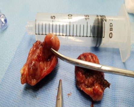
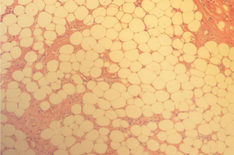
The mass was excised with both tonsils under general anesthesia (Figure 1). The operation went well and the excised mass was sent for histopathology examination, which showed a yellowish mass with a smooth surface measuring 2 × 1 × 1 cm. Low power microscopic examination revealed a well-circumscribed fat tissue composed of lobules and mature adipocytes with characteristic eccentric nuclei and intervening variable thickness bands of fibrous tissue (Figure 2). No cytonuclear atypia or mitotic activity was identified. The postoperative course was uneventful and the patient was discharged after one day. A three months follow-up revealed no evidence of recurrence.
Discussion
Benign neoplasms of the tonsils are extremely rare and typically classified based on the predominant histological tissue type, including squamous papilloma, fibrovascular polyp, lymphangiectatic fibrous polyp, hemangioma, and lipoma [6]. Given their rarity, tonsillar masses may initially raise suspicion for more common pathologies such as chronic tonsillitis, hypertrophic lymphoid tissue, or even malignancies. Therefore, lipoma should be considered in the differential diagnosis of unilateral tonsillar enlargement, particularly when presenting with a soft, slow-growing, non-tender mass.
This report adds to the limited number of documented cases of tonsillar lipoma in the literature [2],[3],[4],[5]. The exact pathogenesis of tonsillar lipomas remains uncertain. However, this case supports the theory proposed by Begin and Frenkiel, who argued that tonsillar lipomas represent true benign neoplasms rather than hamartomatous malformations, based on the absence of other germ cell layers in histopathologic evaluation [5]. Additionally, chronic irritation and local trauma have been suggested as possible contributing factors in their development [2].
Our patient, a 28-year-old male, presented with a foreign body sensation in the throat and was found to have a lipoma arising from the upper pole of the right tonsil. He underwent bilateral tonsillectomy with no recurrence observed after a three-month follow-up. While some reports advocate for conservative excision of the mass alone [3],[5],[7], bilateral tonsillectomy was selected in our case to ensure complete excision, facilitate histopathological diagnosis, and reduce the risk of recurrence. This approach is consistent with the most frequently reported management strategy for tonsillar lipomas in the literature [7].
Tonsillar lipomas may present with a range of clinical symptoms including cough [8], voice change [9], or obstructive sleep apnea [7], although they are frequently discovered incidentally. Most lipomas arise from the tonsillar body, though cases originating from the lower or upper poles have also been described [7], as demonstrated in our patient. The clinical and histopathological findings in this case further underscore the variable presentation and diagnostic challenges of tonsillar lipomas, especially in low-prevalence regions.
Conclusion
Lipoma of the tonsils is a very rare benign tumor that is usually discovered accidentally and my present differently. The current report presents a case of tonsillar lipoma manifested as foreign body sensation in the throat which was managed by bilateral tonsillectomy with no recurrence after a three months follow-up.
REFERENCES
1.
Hyams VJ. Differential diagnosis of neoplasia of the palatine tonsil. Clin Otolaryngol Allied Sci 1978;3(2):117–26. [CrossRef]
[Pubmed]

2.
El-Monem MHA, Gaafar AH, Magdy EA. Lipomas of the head and neck: Presentation variability and diagnostic work-up. J Laryngol Otol 2006;120(1):47–55. [CrossRef]
[Pubmed]
3.
Wang CP, Kwan PC, Ho CY. Lipoma of the palatine tonsil. J Formos Med Assoc 2007;106(8):673–5. [CrossRef]
[Pubmed]
4.
Harada H, Kashiwagi S, Morimatsu M, Kameyama T, Takahashi M. Tonsillar lipoma: A case report. J Laryngol Otol 1995;109(7):662–4. [CrossRef]
[Pubmed]
5.
Nandakumar R, Inchara YK, D’Souza O, Sreenivas V, Mohanty S. Fibrolipoma of the tonsil. Indian J Pathol Microbiol 2010;53(3):562–3. [CrossRef]
[Pubmed]
6.
Bégin LR, Frenkiel S. Polypoid lipoma of the palatine tonsil. J Laryngol Otol 1993;107(6):556–8. [CrossRef]
[Pubmed]
7.
Kanotra SP, Davies J. Management of tonsillar lipoma: Is tonsillectomy essential? Case Rep Otolaryngol 2014;2014:451570. [CrossRef]
[Pubmed]
8.
Moon TH, Lee DJ, Lee SJ, Jung PS. A case of lipoma of epiglottis. Korean J Otorhinolaryngol-Head Neck Surg 2009;52(3):270–2. [CrossRef]
9.
Naruse T, Yanamoto S, Yamada S, Rokutanda S, Kawakita A, Takahashi H, et al. Lipomas of the oral cavity: Clinicopathological and immunohistochemical study of 24 cases and review of the literature. Indian J Otolaryngol Head Neck Surg 2015;67(Suppl 1):67–73. [CrossRef]
[Pubmed]
SUPPORTING INFORMATION
Acknowledgments
During the drafting and refinement of this manuscript, the authors used digital writing tools, specifically, ChatGPT (version GPT-4, OpenAI, San Francisco, CA, USA), to make the language clearer and keep the expression consistent.
The AI tool was used only to help with grammar, sentence structure, and style. Mostly in the Abstract, Introduction, and Discussion sections. No content was generated for clinical data, analysis, or interpretation.
All AI-assisted revisions were carefully reviewed, verified for accuracy, and approved by the authors. Responsibility for the integrity and originality of the manuscript, it remains entirely with the authors.
Lenah Khalid AlFadhil - Conception of the work, Design of the work, Acquisition of data, Drafting the work, Final approval of the version to be published, Agree to be accountable for all aspects of the work in ensuring that questions related to the accuracy or integrity of any part of the work are appropriately investigated and resolved.
Abdulaziz Mohammed AlGhamdi - Conception of the work, Design of the work, Drafting the work, Revising the work critically for important intellectual content, Final approval of the version to be published, Agree to be accountable for all aspects of the work in ensuring that questions related to the accuracy or integrity of any part of the work are appropriately investigated and resolved.
Guaranter of SubmissionThe corresponding author is the guarantor of submission.
Source of SupportNone
Consent StatementWritten informed consent was obtained from the patient for publication of this article.
Data AvailabilityAll relevant data are within the paper and its Supporting Information files.
Conflict of InterestAuthors declare no conflict of interest.
Copyright© 2025 Lenah Khalid Al Fadhil. This article is distributed under the terms of Creative Commons Attribution License which permits unrestricted use, distribution and reproduction in any medium provided the original author(s) and original publisher are properly credited. Please see the copyright policy on the journal website for more information.


